The Bunion Cure Patient Visual Guide
Bunion Surgery Healing: What The X-rays Show Over Time
See how minimally invasive bunionectomy healing can look from before surgery through 8 months, including temporary pin protection, early bone stability, strengthening, and later remodeling.
How To Read This Progression
X-rays show bone position and healing. They do not show exactly how your foot will feel, how much swelling you will have, or when you personally should increase activity.
Your X-rays may look different. Appearance can vary based on bunion severity, whether hammertoes are corrected, the exact procedure performed, bone quality, and how quickly you heal.

The Healing Stages On X-ray

Before surgery
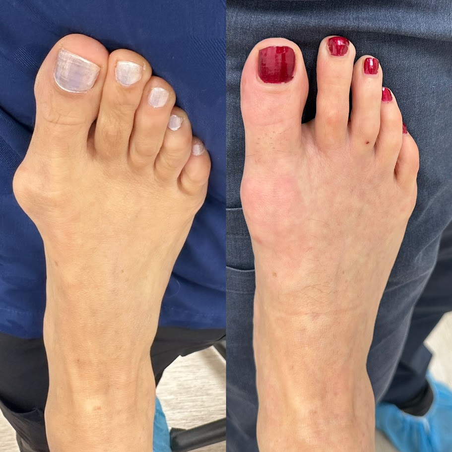
The bunion angle and bump are visible before correction. This is the starting point for surgical planning.

Immediately after surgery
The bone has been repositioned and protected with a temporary pin while early healing begins.

Around 4 weeks
The bone is typically stable by week 3, so the temporary pin is commonly removed at week 4.

Around 8 weeks
The bone keeps getting stronger each week while activity increases carefully.

Around 8 months
The bone continues remodeling after week 12 and becomes more efficient over time.
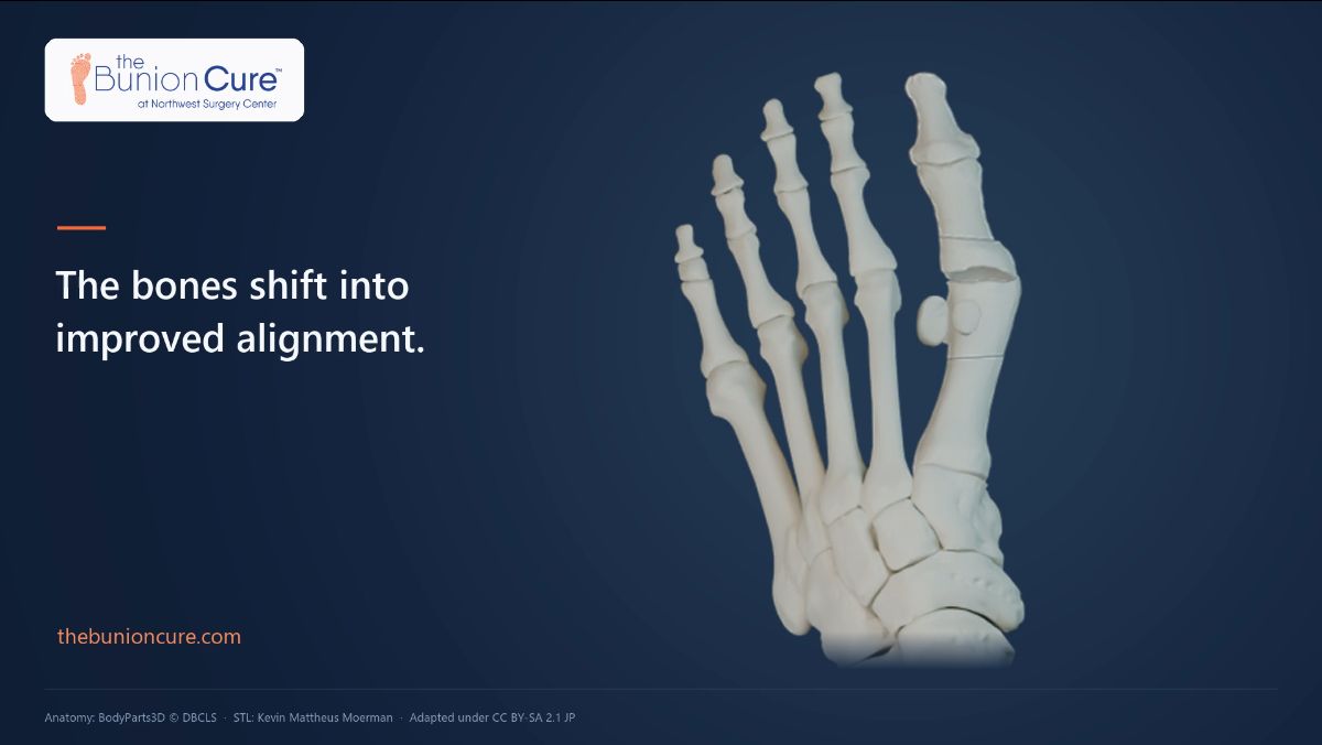
Watch How The Correction Is Made
See how the bone is repositioned and supported with a temporary pin before following the healing stages on real X-rays.
Why A Temporary Pin Is Used
The temporary pin holds the bone while the bone heals. Hardware used for osteotomies or bunion surgery is there to hold the bone in position while healing occurs.
Once the bone is healed, that hardware is no longer needed. Plates and screws can sometimes cause irritation and may need to be removed later. With this approach, the temporary pin is removed around 4 weeks, when the bone is stable, leaving no permanent hardware in your foot.
Why week 4? Bone is typically stable by week 3. The pin is commonly removed at week 4 because the bone is stable intrinsically and the pin is no longer needed.
Temporary pin
Holds the bone while it becomes stable, then comes out in clinic around week 4.
Plates and screws
Can remain after the bone heals and may irritate soft tissues or require removal later.
The goal
Stable healing without leaving permanent hardware behind once the bone no longer needs support.
What X-rays Show vs. What Patients Feel
Your X-rays, swelling, shoe fit, and activity tolerance may improve on different timelines. A good X-ray does not always mean the foot feels completely normal yet.
What your X-rays can show
- Bone position after correction
- Whether the alignment is being maintained
- When the bone is stable enough for pin removal
- How the bone strengthens through week 12 and remodels afterward
What you may still notice day to day
- Swelling can outlast pain and affect shoe fit
- Walking is protected early, not unlimited
- Activity should build gradually even when X-rays look good
- Work demands and footwear can change the return timeline
Recovery At A Glance
Your recovery depends on your job demands, swelling, procedure plan, and healing rate. These are common milestones, but your instructions may be more specific.
Surgery day
You walk out with protected weight bearing in a surgical sandal.
Weeks 1-3
Bandage care and swelling control are the priority. Follow-up visits check the correction.
Week 4
The temporary pin is commonly removed because the bone is usually stable enough on its own.
Weeks 4-8
Many people transition toward roomy athletic shoes or Crocs as swelling allows.
Weeks 10-12+
Bone strength is typically back near normal by week 12. Higher-impact activity builds gradually.
What Keeps Happening After The Pin Comes Out
After the temporary pin is removed, the bone keeps getting stronger each successive week. Bone strength is typically back near normal by week 12, but the bone continues remodeling after that and becomes more efficient over time.
This is why patients can often walk early in a protected surgical sandal, while higher-impact activity still needs to build back gradually as the bone continues to strengthen.
What this means for patients
The X-rays help explain why protected walking can start early, while impact activities still wait until the bone is stronger. The process is not just about the incision; it is about stable bone healing over time.